Guest Post by Marie Svoboda, J. Paul Getty Museum

Tempera on wood, about A.D. 170–200
In 2013, the Antiquities Conservation Department at the J. Paul Getty Museum launched a collaborative study inviting major museums around the world to participate in the study and characterization of the manufacturing practices and material makeup of ancient mummy portraits from Roman Egypt. The primary mission of the project is to increase our understanding of material choices, production and sourcing, as well as understand the techniques employed by ancient artists to produce these artifacts. The project has established a new model for online data sharing among scholars world-wide. Known as the APPEAR: Ancient panel painting, Examination, Analysis and Research project, this Getty initiative encourages a close collaboration of its current team of 49 participating institutions, and the guidance of 11 advisory consultants from different backgrounds such as Egyptology, science, ancient history and the arts. A database developed for this project guides the participants through data entry within specific fields, such as visual observations, analytical results and historic research, to insure consistency. Included in the study is the non-destructive and diagnostic tool of technical imaging which has revealed enormous promise for preliminary material characterization and comparative studies.
Portrait of a Young Woman
The mummy portrait used in the example is that of a Portrait of a Young Woman (81.AP.29). As a painted mummy portrait, it was created to be placed over the face of the deceased and incorporated into the body wrappings. The portrait, which dates to approximately the 1st century AD (based on carbon-14 results) is painted using glue tempera on a sycomore wood panel measuring 34.9 x 21.3 cm, with a thickness of 11.03 mm.
For the APPEAR project, the mummy portraits are typically imaged using visible light at full spectra and narrow band passes, ultraviolet radiation 365 nm (UV), near and far infrared at 904 and 1600 nm (IR), visible induced infrared luminescence (VIL) and X-radiography. Each method provides very unique information and comparison between them can help characterize ancient materials, the artist’s working method, damage, modern restoration and repairs and other features not visible to the naked eye.
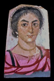
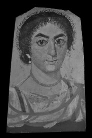
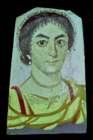
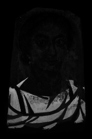
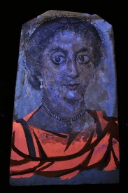
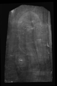
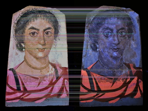
Image Alignment and Visualization
By aligning and precisely stacking the imaging modalities, it is possible to compare the images in detail and better understand the acquired data. To do this, the images were first aligned and resized to a common reference frame such that each pixel in each image aligns exactly to the equivalent pixel in every other imaging modality.
For the examples here, each image was warped in order to perfectly align to the color image taken under visible light. This was carried out using traditional image alignment techniques involving the identification of common key points through feature detection, the calculation of a suitable geometric transform and the warping of the image using this transform. Image processing for this was carried out by IIPImage’s main developer, Ruven, and implemented using Python and OpenCV.
Once aligned, the IIPImage viewer, iipmooviewer, was used to provide not only the ability to interactively zoom and navigate within the high resolution images, but also to compare each of the different technical images. The viewer allows the user to either switch between stacked images or slowly fade between any 2 images via the control panel on the right, thereby allowing the user to compare the two simultaneously and providing an innovative and valuable method of study. The user can select any two images to compare from the drop down menus, and use the slide bar to fade from one image to the other to allow pixel-wise comparison of the material’s photometric response under the given combination of illuminant and filter.

Analyzing the Portrait
The images selected for comparison were full spectrum visible, narrow band visible at 555nm, UV induced visible fluorescence, IR using a 904 nm band-pass filter, x-ray and false color infrared (FCIR).
Imaging Methods
- Color visible image using a modified Nikon D90 camera with a Peca 916 filter and the Mini-CrimeScope 400 unfiltered light source
- Infra-red reflected image (IR) using a modified Nikon D90 camera with a Peca 904 bandpass filter, illuminated with halogen light sources. Carbon black used for the hair, eyes, eyebrows and mouth are visible
- Infra-red false color image (FCIR) created by combining the visible and infrared images and rearranging the color channels in Adobe PhotoShop. The presence of indigo is represented as magenta, seen on the clavus and the folds of her tunic, pale yellow identifies the madder lake and the red iron based pigment appears a darker golden yellow
- 555 nm Image capture at a narrowband pass, using a modified Nikon D90 camera with Peca 916 and a red filter, illuminated with the Mini-CrimeScope 400, filtered at 555 nm. Processed as a monochromatic image. The organic pigment on the tunic luminescences
- Ultra-violet image using a Canon Rebel EOS and Peca 916 filter, illuminated by a Wildfire Long throw series UV light source at 365 nm. The pink tunic exhibits the typical orange/pink fluorescence characteristic of organic lake/madder. The necklace, earrings, whites of her eyes and sketch-like details on her face exhibit a bright fluorescence suggesting the use of a calcium based white
- X-ray, at 28 KV 6.25 mA 2.5 foc –2 minute exposure. There is no evidence for the use of a lead based pigment which would appear as white (opaque) details. Material identifications were corroborated through scientific analysis
Although the x-ray image was included, it does not reveal distinctive features such as radio opaque pigments (e.g., lead white) here. However, this in itself is informative as most portraits use lead pigments. The image generated using VIL, a method of capturing IR luminescence of materials, specifically the ancient synthetic pigment Egyptian blue, was not compared because this material was not identified on this portrait.
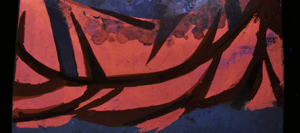
Imaging with ultraviolet radiation highlights the bright orange-pink fluorescence characteristic of the organic pigment madder lake used for her brightly colored garment. In addition to being corroborated analytically, the visible image captured with a narrow band-pass filter at 555nm also helps characterizes the use of the organic lake pigment.
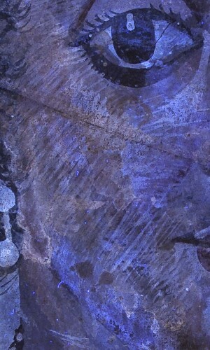
The fine painting technique, such as the cross-hatching brush strokes on her proper right cheek are also enhanced with UV. Other visible features include uneven staining on her face and traces of resin and textile impressions at the perimeter of the panel. When comparing this image with the FCIR image, the use of madder (appearing yellow), iron based pigments (appearing golden yellow/green) and indigo (magenta) are revealed on her garment. Iron based pigment and indigo are also characterized on the clavi (dark vertical stripes on her tunic) and to indicate the folds on her tunic, highlighting the use and complexity of color choices.
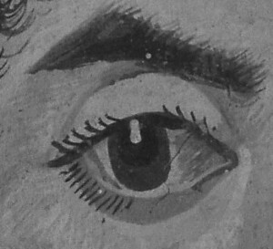
The identification of carbon black is made possible due to the fact that it absorbs infrared radiation and thus appears dark with IR imaging, even when present under paint layers. This technique is typically used for identifying underdrawing on paintings. On the portrait of a woman, carbon black is observed in the hair and various highlight details (eyes and eyelashes). It was also likely mixed with other colors to darken them, such as in the shadows on her neck and clavus. It can be inferred that the order in which the garment was painted is: the ground layer, the tunic, clavus followed by mantle (shawl) because in visible light the proper left clavus is hidden but visible through the paint layers in IR. Through comparison of these various imaging techniques, an intentional color palette and technique begins to emerge and the ancient artist’s methodology is revealed.
Portrait of a Bearded Man

Tempera on wood. A.D. 220–250
A second object chosen for multi-modal image comparison was the Mummy Portrait of a Bearded Man (79.AP.142). This portrait, painted with tempera on a cedar of Lebanon panel, depicts a bearded man holding a glass with wine in his proper right hand and a rose petal garland in his left. The wooden panel measuring 34 × 25 cm and is 1.2 cm thick dates to the first century BC. However, the portrait was stylistically dated to the third century AD, possibly suggesting that the wooden panel was repurposed from another artifact. The same technical image series as with the portrait of a young woman, was selected for use with the IIPImage viewer: full spectrum visible, narrow band visible at 555nm, UV induced visible fluorescence, IR using a 904 nm bandpass filter, X-radiograph and false color infrared (FCIR). The pigments selected to paint the portrait of a bearded man can be characterized through these images and by comparing them. The bright orange fluorescence of madder lake is seen with ultraviolet radiation, in his face, the wine, rose garland and decorative embroidery on the neck-line of his chiton. The identification of madder was compared and further corroborated by its characteristic luminescence when imaged at 555 nm. The X-radiograph shows that details of the bearded man’s face, such as his chin, were painted with a radio opaque pigment, confirmed through analysis as lead white. Comparing the x-ray with the UVF image shows that the artist used lead white to suggest the light colored flesh surrounding the eyes, but the ruddy color of the cheeks were achieved with madder. The infrared image indicates the use of carbon black for his hair, eyes and the details of the wine glass and rose garland. Lastly, the use of indigo to produce the blue clavi, details of the wine glass and combined with a yellow pigment to create the green leaves at the bottom of the rose garland can be identified by the magenta enhancement in the FCIR image. Comparing these images is a non-destructive and effective way to study and reveal the artist’s working methodology, observed through the choice of pigments used and painting techniques.
Special acknowledgement goes to the UCLA / Getty conservation training program for the use and guidance in using the Mini CrimeScope for imaging this mummy portrait and to the Los Angeles County Museum of Art for imaging assistance.
